Anatomage

Login to Anatomage Cloud. Upload, view and send 3D DICOM medical images. Any doctor can easily send a medical image for instant review to another doctor. The recipient can access the image from anywhere and instantly review the image on any device - including mobile.
The Anatomage is a virtual dissection software that visualizes the anatomy systems of human and animal cadavers in 3D. The rich virtual library contains female and male 3D gross anatomy, 3D regional anatomy, and 120 pathological examples. Additionally, the Anatomage contains radiology features such as medical CT, CBCT, and MRI scans. The software is capable of opening and rendering any medical imaging data into an interactive 3D structure. Utilizing the expansive content library, instructors can create customized lecture content to fit their curriculum.
Related Music Albums:.Raaz (2002) songs downloadBollywood Hindi Movie Raaz (2002) songs Download and listen online. Raaz mp3 download. Cast and crew of this hindi movie is Dino Morea, Bipasha Basu, Malini Sharma, Ashutosh Rana. Release date is 1 February 2002.
Find out more about the Anatomage here: https://www.anatomage.com/
To use the anatomage, please email canvas@unthsc.edu to make an appointment.
CardioSource WorldNews Given the myriad of issues surrounding cadaveric dissection as well as the pace of technology change, a number of innovative new tools have been released to enable virtual anatomy education. I recently came across a leading platform called Anatomage at the AMEE International Conference for Medical & Health Education in Barcelona this August and took the opportunity to speak with their team about the technology and its applications.
How did Anatomage get started?
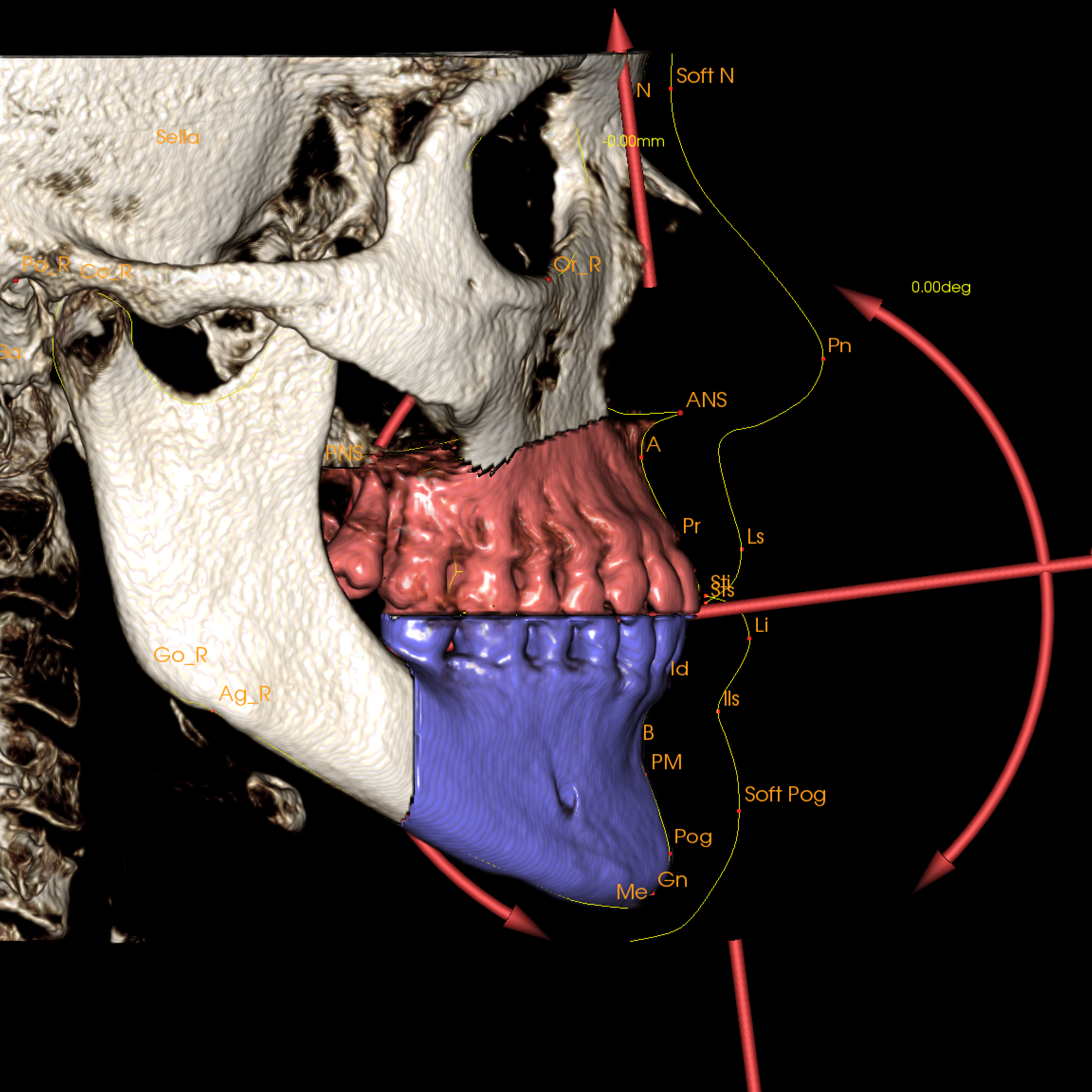
Anatomage was founded in 2004. Anatomage developed the first volume reconstruction software for the dental market. The flagship product for the company was InVivoDental. The InvivoDental software takes in cone-beam CT scan files and renders them into a 3D image. The InvivoDental software is used for orthodontic simulations and implant surgery treatment planning. In 2011 the Anatomage Table was released as a platform to present anatomy in a life size scale. We used the same technology to load in CT and MRI scans and render them in 3D to create a Table that allows for visualization of the human body. The Table was designed to resemble the form factor of a dissection table.
Can you explain the technology underlying your products?
The Table’s content consists of two distinctive sides. The first is displaying male and female gross anatomy, primarily used in anatomy education. The second is being able to load in any CT or MRI scan to view it in a fully three dimensional perspective. The cadavers on the Table are taken from the Visible Korean project. The cadavers were flash frozen at the time of the death and then sliced axially at 0.2 mm sections. We then went through and segmented out each structure of the body to create a fully interactive cadaver. We have a clinical library of over a 1,000 cases that range from rare and unique pathology to more common surgical procedures and normal anatomy. It also contains animals scans, embryology, and histology.
What are the current ways Anatomage is used?
The Anatomage Table is being used by hundreds of institutions around the world. The Table is primarily used for anatomy education and it is being used by every kind of organization from high schools to medical schools. Each institution has a unique way of incorporating the Table into their curriculum. The Table allows a student to redo any cut or any mistake that could be detrimental when working directly on a cadaver. There is an ease of use with loading up a portion of the body and clicking on a structure to get it’s annotation. Teachers can create material like lectures or quizzes directly on the Table and save it for later use. Teaching hospitals, simulation centers and research facilities all use the Table. Patient scans can be loaded directly onto the Table for visualization in 3D. The Anatomage Table has been cleared by the FDA for radiologists, clinicians, referring physicians and other qualified individuals to retrieve, process, render, review, and assist in diagnosis.
Please describe how cardiologists and practicing physicians may use your products.
Clinicians are using the Table as a platform for patient consultation, department collaboration, and assistance in diagnosis. Clinicians can use the Table to teach their patients about surgery planning and demonstrate the importance of the procedure they are receiving. Some facilities are using the Table’s complimentary software to create personalized 3D prints of the patient’s CT scan to give the patient a better understanding of the disease that they are facing. Teams of clinicians from different departments have used the Anatomage Table as a point of collaboration to discuss the best way to treat a patient. Radiologists and physicians have worked side by side to compare the 2D images from a CT scan to the 3D image created by the Table to devise a pathway for a surgical procedure. The Table is able to assist the overall diagnosis process and provide a better method for surgeons to plan their surgical procedures.
Where do you see Anatomage in 5 years? Windows 8 end of support. 15 years?
In 5 years, we see Anatomage continuing to be the premiere software and hardware provider for clinicians, health care services, and medical education. We strive to be on the cutting edge and we position ourselves to be the go-to technology when it comes to advanced education and medical applications. Whether it is virtual dissection, medical diagnosis, or surgical planning, we want to be the number one solutions provider. Our technology has been adopted in thousands of institutions worldwide and we aim to be in hundreds of thousands more. In 15 years, we endeavor to create even newer cutting-edge technology that we hope will revolutionize not only medical education, but also medical practice and health care services. We want to create technology that is both practical and revolutionary. As a medical device company, we understand that medicine is an ever growing and developing science, and we want to be at the very forefront of creating new innovative technology that the field and industry find effective and will widely adopt.
Shiv Gaglani is an MD/MBA candidate at the Johns Hopkins School of Medicine and Harvard Business School. He writes about trends in medicine and technology and has had his work published in Medgadget, The Atlantic, and Emergency Physicians Monthly.
| Read the full September issue of CardioSource WorldNews at ACC.org/CSWN |
Clinical Topics:Invasive Cardiovascular Angiography and Intervention, Noninvasive Imaging, Interventions and Imaging, Magnetic Resonance Imaging
Keywords:CardioSource WorldNews, Imaging, Three-Dimensional, Magnetic Resonance Imaging, Software, Cone-Beam Computed Tomography
< Back to Listings
